Product Information
- Product Type
- Monoclonal Antibody
- Clone Number
- AT17D10
- UniProt No.
- P09104
- NCBI Accession No.
- NP_001966
- Alternative names
- Enolase 2 (gamma, neuronal), ENO2, NSE, Neuron-Specific Enolase, 2 phospho D glycerate hydrolyase, Eno 2, ENOG, Enolase 2 gamma neuronal, Enolase2, Gamma enolase, Neural enolase, Neuron specific enolase, Neuron specific gamma enolase, Neurone specific enolase.
Product Specification
- Host
- Mouse
- Reacts With
- Human
- Concentration
- 1mg/ml (determined by BCA assay)
- Formulation
- Liquid in. Phosphate-Buffered Saline (pH 7.4) with 0.02% Sodium Azide, 10% glycerol
- Immunogen
- Recombinant human NSE (1-434aa) purified from E. coli
- Isotype
- IgG2b kappa
- Purification
- By protein-A affinity chromatography
- Applications
- ELISA, WB, ICC/IF
- Usage
- The antibody has been tested by ELISA, Western blot analysis to assure specificity and reactivity. Since application varies, however, each investigation should be titrated by the reagent to obtain optimal results. Recommended dilution range for Western blot analysis is 1:500 ~ 1:5000. Recommended starting dilution is 1:1000.
- Storage
- Can be stored at +2C to +8C for 1 week. For long term storage, aliquot and store at -20C to -80C. Avoid repeated freezing and thawing cycles.
Data
Immunocytochemistry/Immunofluorescence (ICC/IF)
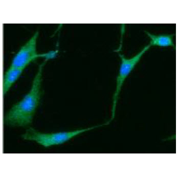
ICC/IF analysis of NSE in U87MG cells line, stained with DAPI (Blue) for nucleus staining and monoclonal anti-human NSE antibody (1:100) with goat anti-mouse IgG-Alexa fluor 488 conjugate (Green).
Western blot analysis (WB)
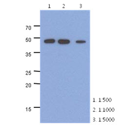
The extracts of mouse brain (40ug) were resolved by SDS-PAGE, transferred to PVDF membrane and probed with anti-human NSE antibody (1:500 ~ 1:5000). Proteins were visualized using a goat anti-mouse secondary antibody conjugated to HRP and an ECL detection system.
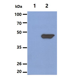
The Cell lysates (5ug) were resolved by SDS-PAGE, transferred to PVDF membrane and probed with anti-human NSE antibody (1:1000). Proteins were visualized using a goat anti-mouse secondary antibody conjugated to HRP and an ECL detection system.
Lane 1.: 293T cell lysate
Lane 2.: NSE Transfected 293T cell lysate
Lane 1.: 293T cell lysate
Lane 2.: NSE Transfected 293T cell lysate
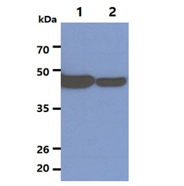
The Cell lysates (20ug) were resolved by SDS-PAGE, transferred to PVDF membrane and probed with anti-human NSE antibody (1:1000). Proteins were visualized using a goat anti-mouse secondary antibody conjugated to HRP and an ECL detection system.
Lane 1.: U-87 MG cell lysate
Lane 2.: Jurkat cell lysate
Lane 1.: U-87 MG cell lysate
Lane 2.: Jurkat cell lysate
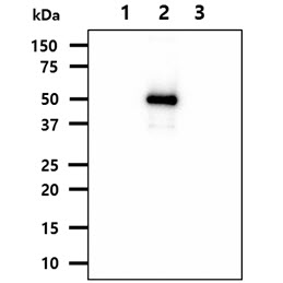
The recombinant proteins (50ng) were resolved by SDS-PAGE, transferred to PVDF membrane and probed with anti-human NSE antibody (1:1000). Proteins were visualized using a goat anti-mouse secondary antibody conjugated to HRP and an ECL detection system.
Lane 1.: Recombinant protein ENO1
Lane 2.: Recombinant protein NSE (ENO2)
Lane 3.: Recombinant protein ENO3
Lane 1.: Recombinant protein ENO1
Lane 2.: Recombinant protein NSE (ENO2)
Lane 3.: Recombinant protein ENO3
Note: For research use only. This product is not intended or approved for human, diagnostics or veterinary use.