Product Information
- Product Type
- Monoclonal Antibody
- Clone Number
- AT10F1
- UniProt No.
- P61956
- NCBI Accession No.
- NP_008868
- Alternative names
- ubiquitin like protein SMT3B.Small ubiquitin-related modifier 3, SuMO3, SuMO2, SMT3H2, SMT3H1, SMT3B, SMT3A, SMT3 suppressor of mif two 3 homolog 2, SMT3 homolog 2, SMT 3B, Small ubiquitin-related modifier 2 SMT3 suppressor of mif two 3 homolog 2 (S. cerevisiae), Small ubiquitin related modifier 2, Small ubiquitin like modifier 2, Sentrin2, MGC117191, HSMT3
Product Specification
- Host
- Mouse
- Reacts With
- Human
- Concentration
- 1mg/ml (determined by BCA assay)
- Formulation
- Liquid in. Phosphate-Buffered Saline (pH 7.4) with 0.02% Sodium Azide, 10% glycerol
- Immunogen
- Recombinant human SUMO2 (1-93aa) purified from E.coli
- Isotype
- IgG2b kappa
- Purification
- By protein-G affinity chromatography
- Applications
- ELISA, WB, ICC/IF, IHC, FACS
- Usage
- The antibody has been tested by ELISA, Western blot, ICC/IF, FACS and IHC analysis to assure specificity and reactivity. Since application varies, however, each investigation should be titrated by the reagent to obtain optimal results.
- Storage
- Can be stored at +2C to +8C for 1 week. For long term storage, aliquot and store at -20C to -80C. Avoid repeated freezing and thawing cycles.
Data
Immunocytochemistry/Immunofluorescence (ICC/IF)
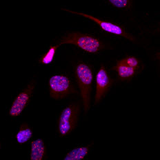
Immunofluorescence of human HeLa cells stained with Hoechst 3342 (Blue) for nucleus staining and monoclonal anti-human SUMO2/3 antibody (1:500) with Texas Red (Red).
Western blot analysis (WB)
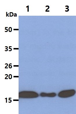
The cell lysates(40ug) were resolved by SDS-PAGE, transferred to PVDF membrane and probed with anti-human SUMO2/3 antibody (1:1000). Proteins were visualized using a goat anti-mouse secondary antibody conjugated to HRP and an ECL detection system.
Lane 1.: HeLa cell lysate
Lane 2.: Jurkat cell lysate
Lane 3.: K562 cell lysate
Lane 1.: HeLa cell lysate
Lane 2.: Jurkat cell lysate
Lane 3.: K562 cell lysate
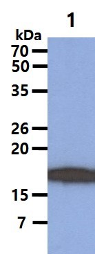
The cell lysate(40ug) was resolved by SDS-PAGE, transferred to PVDF membrane and probed with anti-human SUMO2/3 antibody (1:1000). Proteins were visualized using a goat anti-mouse secondary antibody conjugated to HRP and an ECL detection system.
Lane 1.: HL-60 cell lysate
Lane 1.: HL-60 cell lysate
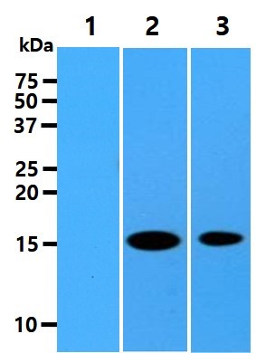
The recombinant proteins (10ng) were resolved by SDS-PAGE, transferred to PVDF membrane and probed with anti-human SUMO2/3 antibody (1:1000). Proteins were visualized using a goat anti-mouse secondary antibody conjugated to HRP and an ECL detection system.
Lane 1.: Recombinant human SUMO1 protein
Lane 2.: Recombinant human SUMO2 protein
Lane 3.: Recombinant human SUMO3 protein
Lane 1.: Recombinant human SUMO1 protein
Lane 2.: Recombinant human SUMO2 protein
Lane 3.: Recombinant human SUMO3 protein
Flow cytometry (FACS)
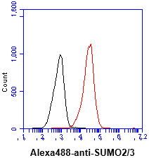
Flow cytometry analysis of SUMO2/3 in jurkat cell line, staining at 2-5ug for 1x10^6cells (red line). The secondary antibody used goat anti-mouse IgG Alexa fluor 488 conjugate. Isotype control antibody was mouse IgG (black line).
IHC Comment And Pic
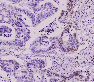
Paraffin embedded sections of colorectal cancer tissue were incubated with anti-human SUMO2/3 antibody (1:50) for 2 hours at room temperature. Antigen retrieval was performed in 0.1M sodium citrate buffer and detected using Diaminobenzidine (DAB).
Note: For research use only. This product is not intended or approved for human, diagnostics or veterinary use.