Product Information
- Product Type
- Monoclonal Antibody
- Clone Number
- 3B6
- UniProt No.
- P37840, Q16143
- NCBI Accession No.
- NP_000336, NP_003076
- Alternative names
- SNCA, NACP, PARK1, PARK4, PD1, α-synuclein Non-A beta component of AD amyloid, Non-A4 component of amyloid precursor, Parkinson disease 4, autosomal dominant Lewy body
Product Specification
- Host
- Mouse
- Reacts With
- Human
- Concentration
- 1mg/ml (determined by BCA assay)
- Formulation
- Liquid in. Phosphate-Buffered Saline (pH 7.4) with 0.02% Sodium Azide, 10% glycerol
- Immunogen
- Recombinant human a-synuclein (119-140aa) purified from E. coli
- Isotype
- IgG1 kappa
- Purification
- By protein-G affinity chromatography
- Applications
- ELISA, WB, ICC/IF, FACS
- Usage
- The antibody has been tested by ELISA, Western blot, ICC/IF and FACS analysis to assure specificity and reactivity. Since application varies, however, each investigation should be titrated by the reagent to obtain optimal results.
- Storage
- Can be stored at +2C to +8C for 1 week. For long term storage, aliquot and store at -20C to -80C. Avoid repeated freezing and thawing cycles.
Data
Immunocytochemistry/Immunofluorescence (ICC/IF)
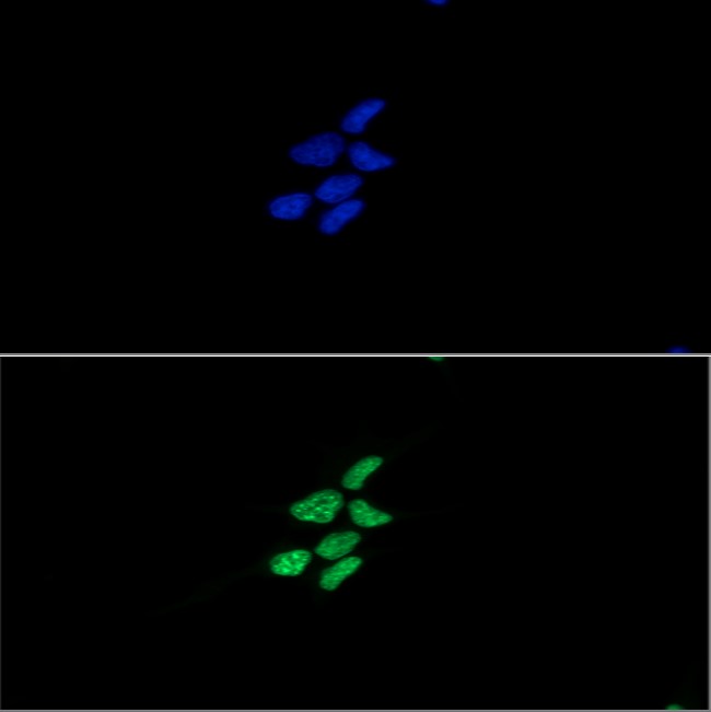
ICC/IF analysis of alpha, beta-Synuclein in LNCaP cells. The cell was stained with ACS0632 (1:100). The secondary antibody (green) was used Alexa Fluor 488. DAPI was stained the cell nucleus (blue).
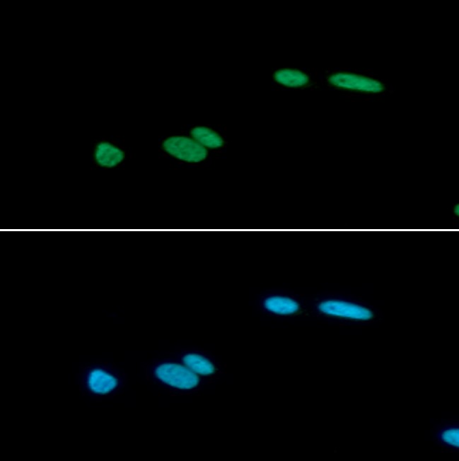
ICC/IF analysis of alpha, beta-Synuclein in U87MG cells. The cell was stained with ACS0632 (1:100). The secondary antibody (green) was used Alexa Fluor 488. DAPI was stained the cell nucleus (blue).
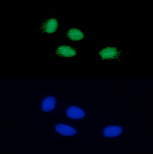
ICC/IF analysis of alpha, beta-Synuclein in C6 cells. The cell was stained with ACS0632 (1:100). The secondary antibody (green) was used Alexa Fluor 488. DAPI was stained the cell nucleus (blue).
Western blot analysis (WB)
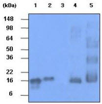
The recombinant protein (20ng) and tissue lysates (30ug) were resolved by SDS-PAGE, transferred to PVDF membrane and probed with anti-human HSP60 antibody (1:1000). Proteins were visualized using a goat anti-mouse secondary antibody conjugated to HRP and an ECL detection system.
Lane 1.: Recombinant human alpha-synuclein protein
Lane 2.: Recombinant human beta-synuclein protein
Lane 3.: Recombinant human gamma-synuclein protein
Lane 4.: Mouse brain Tissue lysate
Lane 5.: Rat brain Tissue lysate
Lane 1.: Recombinant human alpha-synuclein protein
Lane 2.: Recombinant human beta-synuclein protein
Lane 3.: Recombinant human gamma-synuclein protein
Lane 4.: Mouse brain Tissue lysate
Lane 5.: Rat brain Tissue lysate
Flow cytometry (FACS)
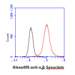
Flow cytometry analysis of alpha, beta-Synuclein in LNCaP cell line, staining at 2-5ug for 1x10^6cells (red line). The secondary antibody used goat anti-mouse IgG Alexa fluor 488 conjugate. Isotype control antibody was mouse IgG (black line).
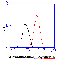
Flow cytometry analysis of alpha, beta-Synuclein in U87MG cell line, staining at 2-5ug for 1x10^6cells (red line). The secondary antibody used goat anti-mouse IgG Alexa fluor 488 conjugate. Isotype control antibody was mouse IgG (black line).
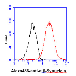
Flow cytometry analysis of alpha, beta-Synuclein in C6 cell line, staining at 2-5ug for 1x10^6cells (red line). The secondary antibody used goat anti-mouse IgG Alexa fluor 488 conjugate. Isotype control antibody was mouse IgG (black line).
Note: For research use only. This product is not intended or approved for human, diagnostics or veterinary use.